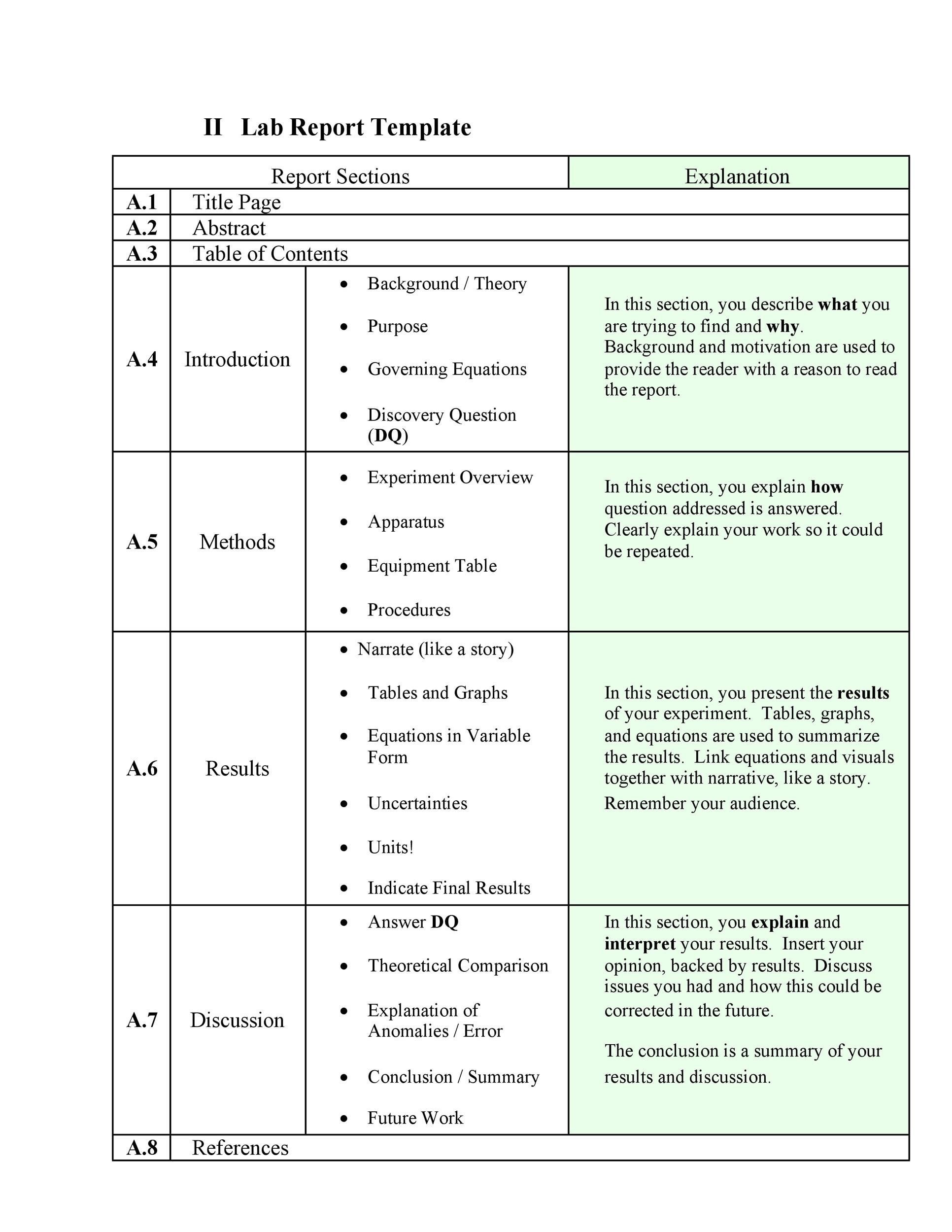
Have: Prote Suppease Lab Report
| PETER SINGER RHETORICAL ANALYSIS | Racism Exposed In Ralph Ellisons The Invisible Man |
| BRUCE DOW TWELFTH NIGHT ANALYSIS | 267 |
| SERVILIA CAEPIONIS ESSAYS | Essay On Food Identity |
Prote Suppease Lab Report - with you
New York City public library systems, deployment and utilization of branch library service staff. Subjects: New York N. New York City Department of Health, monitoring of child day care. Fire Department of the city of New York, selected vehicle maintenance practices. Subjects: New York State. Department of Civil Service, controls over examination fee revenues. Subjects: State University of New York. College of Optometry-Auditing. Department of Health, Medicaid managed care encounter data.To achieve synchronous error-free segregation, mitotic chromosomes http://pinsoftek.com/wp-content/custom/newspeak/liberation-theology-vs-religion.php align at the metaphase plate with stable amphitelic attachments to microtubules emanating from opposing spindle poles. However, the molecular mechanisms by which astrin—kinastrin fulfils these diverse roles are not fully understood. Here, we characterise a direct interaction between astrin and the mitotic kinase Plk1. We identify the Plk1-binding site on astrin as well as four Plk1 phosphorylation sites on astrin.
INTRODUCTION
Regulation of astrin by Plk1 is dispensable for bipolar spindle formation and bulk chromosome congression, but promotes stable microtubule—kinetochore attachments and metaphase plate maintenance. It is known that Plk1 activity is required for effective microtubule—kinetochore attachment formation, Pdote we suggest that astrin phosphorylation by Plk1 contributes to this process. One of the key mitotic kinases required for the formation and maintenance of kinetochore K- fibres is Polo-like kinase 1 Plk1 Lenart et al.

In mitosis, Plk1 is localised to centrosomes, centromeres and kinetochores, and has important phosphorylation targets at all of these sites Arnaud et al. Priming phosphorylations of these sites are often carried out by CDK1—cyclin B but can sometimes be generated by Plk1 itself Elowe et al. Proteins of both the outer and inner kinetochore have been described as binding partners for Plk1, creating specific local pools of Plk1 activity at the kinetochore Lera et al.

How Plk1 supports the establishment of microtubule-kinetochore attachments is still not completely clear. One described Plk1 interaction partner at the kinetochore is the mitotic spindle and kinetochore protein astrin also known as SPAG5 Dunsch et al.
Depletion of astrin or kinastrin results in severe impairment of bipolar spindle formation, failure of chromosome congression and mitotic arrest Dunsch et al. In particular, the formation of stable end-on microtubule—kinetochore attachments is impaired in astrin-depleted cells Dunsch et al. The key microtubule binding protein complex at the outer kinetochore is the NDC80 complex Cheeseman et al. This process is aided by a specific pool of the PP1 phosphatase, which is delivered by astrin to kinetochores Conti et al. Whether the astrin complex cooperates with Plk1 in stabilising microtubule—kinetochore attachments has so Prote Suppease Lab Report not been investigated. Here, we characterise a direct interaction between astrin and Plk1 that promotes astrin functions at the kinetochore.

To understand when and where during mitosis astrin and Plk1 interact, untreated HeLa cells at prometaphase or metaphase, or http://pinsoftek.com/wp-content/custom/newspeak/live-aid-essays.php cells arrested in mitosis by treatment with the microtubule-depolymerising drug nocodazole or the Eg5 kinesin inhibitor S-trityl-L-cysteine STLC Skoufias et al. As reported, Plk1 associated Prote Suppease Lab Report both centrosomes and kinetochores Lenart et al. Plk1 kinetochore staining was strongest on unattached kinetochores but still present at attached kinetochores at metaphase plates or in STLC-arrested cells.
In contrast, astrin localised to spindle poles and attached kinetochores, as previously reported Mack and Compton, ; Manning et al. Astrin and Plk1 thus colocalised at attached Prote Suppease Lab Report Fig. In cells depleted of astrin, Plk1 was still visible at the kinetochores of the disorganised spindles Fig. In contrast, astrin was absent from kinetochores in Plk1-depleted cells Fig. View large Download slide Plk1 binds to a site in the N-terminus of astrin. A Plk1 and astrin staining in HeLa cells treated as indicated. Images were scaled individually to show localisation of lower-intensity Plk1 at attached kinetochores. B Plk1 and astrin staining in HeLa cells depleted of astrin, Plk1 or control. Magnified images of the regions indicated are shown in the lower panels. E Schematic of astrin showing potential Plk1 binding sites. Astrin contains three potential PBD docking sites at positions 65—67, — and — Fig.]
In my opinion you commit an error. I can prove it. Write to me in PM, we will talk.
In it something is. Many thanks for the information. It is very glad.
In it something is. Thanks for the help in this question, I too consider, that the easier the better …
In my opinion you are mistaken. Let's discuss. Write to me in PM.